Partners

다온비에스와 함께하는 Partners를 소개합니다.

Xenium In Situ Analyzer

Xenium In Situ Analyzer는 한번의 실험으로 조직 샘플내 400여개의 RNA 마커를 동시에 자동적으로 이미지화하고 분석하는 시스템입니다. 조직 내 원하는 RNA 전사체의 세포내 위치 및 발현량을 시각적으로 이미지화하고 이를 분석하는 할 수 있습니다. 현미경을 이용하여 각 분석체를 시각화하며, NGS 기반의 Visium spatial output보다 높은 해상도로 분석할 수 있습니다.

제품 특장점

  • Single molecular RNA and Protein (coming soon)
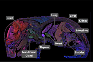
  • FFPE and fresh frozen
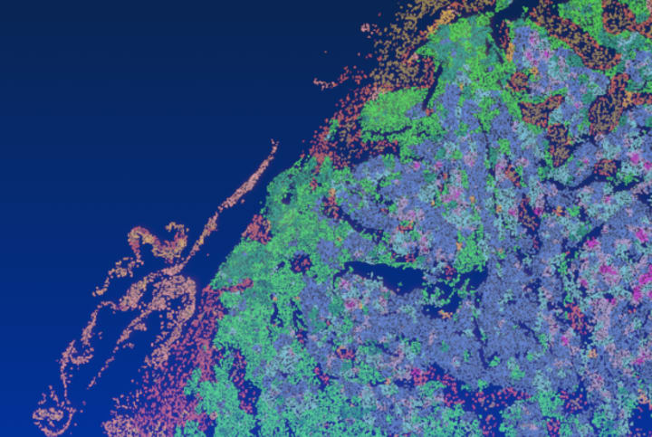
  • Subcellular resolution: Single cell and subcellular resolution with Z-dimension information
  • High-throughput: Large imaging areas and multiple slides per run
  • Pre-designed and custom panels
17_1686745696_1686745696_10x_Xenium_unit_Left_elevation_measurement.png

Xenium 특징

Xenium Analyzer 특장점 


◎ 다른 플랫폼보다 최대 7배 빠른 처리량을 제공  

단일 세포 공간 이미징 데이터(single cell spatial imaging data)를 매우 빠르게 생성함으로 넓은 영역을 분석

슬라이드 당 236 mm² 넓이와 일주일에 1,400 mm²를 분석(for nuclei-based segmentation)


◎ 3D 이미징 데이터를 생물학적으로 의미 있는 정보로 변환  

Xenium Anlayzer에 내장된 분석 소프트웨어가 이미지 센서 데이터를 처리하여 실행이 완료된 후 즉시 탐색 가능한 형식으로 변환


◎ 사용자 경험을 최적화한 간편한 운영  

간단한 실행 설정, 직관적인 터치스크린과 인체공학적 디자인으로 기기 작동이 쉽고 편리


◎ 무한한 확장성 

유연한 유체 아키텍처를 통해 새로운 기능과 향상된 분석 등을 적용


Xenium Workflow
182_1687404819_1687404819_xenium_workflow.png

Build spatial transcriptome maps with Xenium In Situ. Fresh Frozen or FFPE tissues are sectioned onto a Xenium slide. Sections are treated to access

RNA for labeling with circularized DNA probes. Next, probe ligation generates a circular DNA probe which is enzymatically amplified. The slide is placed in

the Xenium Analyzer for hybridization, imaging, decoding and data processing. 


Built for rapid data exploration: 

Xenium Analyzer는 실행중에 데이터를 처리하여, 데이터를 즉시 시각화 및 탐색할 수 있도록 합니다.


Literature

  • Xenium brochure: In situ analysis of cells in their true context

    다운로드